Guttae can occur on the basement membrane of the cornea, which carries endothelial cells. Endothelial cells are located on the back surface of the cornea and are responsible for actively pumping out fluid and maintaining the cornea's transparency. In the presence of many guttae, the endothelial cells lose their function and cannot hold the cornea's transparency in the long run. This often leads to irreversible visual impairment, sometimes so severe that a new cornea's transplantation becomes necessary. Guttae sometimes remain undetected in the preoperative routine inverted mirror microscopy.
The goal of KIttata (Artificial Intelligence for the Detection and Classification of the Corneal Guttata in the Corneal Bank prior to Keratoplasty) is to develop an AI classification algorithm that predicts the transplantation suitability of a donor cornea. This is intended to increase the long-term survival rate of corneal transplants, reduce the need for retransplantation, and reduce the associated health care costs.
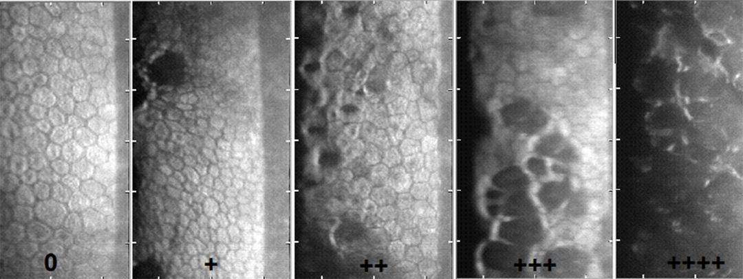
The probability of the existence of undetected guttae in a corneal graft depends on morphological criteria such as differences in cell color, irregularities in cell shape, presence of vesicles, deformities of the cell membrane, or presence of areas without cells.
"While our Ph.D. student Tarek Safi was establishing morphological criteria for the detection of masked guttae on the donor cornea in the corneal bank, he came up with the idea of using artificial intelligence for the detection of alarming structural abnormalities. AI is currently finding many applications in many areas of medicine, including ophthalmology. By using this advanced technology as image analysis and recognition tool, a milestone in the quality assurance of corneal donor tissue could be achieved," explains Prof. Dr. Berthold Seitz, Director of the Department of Ophthalmology at Saarland University Hospital.
Scientists from the DFKI research area Cognitive Assistants use AI methods to develop procedures that implement these criteria. This is essentially a classification algorithm that uses the parameters resulting from the operations to predict whether a given donor cornea is healthy. They use microscopically acquired image data of the corneas, which represent these details, as input parameters for an AI-supported evaluation algorithm (classifier).
"Our goal is to optimize the classification of corneas and achieve more accurate results than the methods used so far. To achieve this, we use a deep learning method, i.e., machine learning algorithms based on complex neural networks. For this purpose, neural models are created that are derived from information from the corneal bank, i.e., images and parameters," says DFKI project manager Dr. Jan Alexandersson.
Finally, the results are evaluated by a clinical expert. This assessment serves as a basis for adjusting the models' input parameters and helps to optimize the model step by step.
Projektinfos
Sponsor: Dr. Rolf M. Schwiete Foundation, Mannheim
Volume: approx. 215,000 €.
Duration: 1 year
Partner: German Research Center for Artificial Intelligence - DFKI, Saarbrücken
Saarland University Hospital - Department of Ophthalmology, Homburg